

 Created by gudegnaw
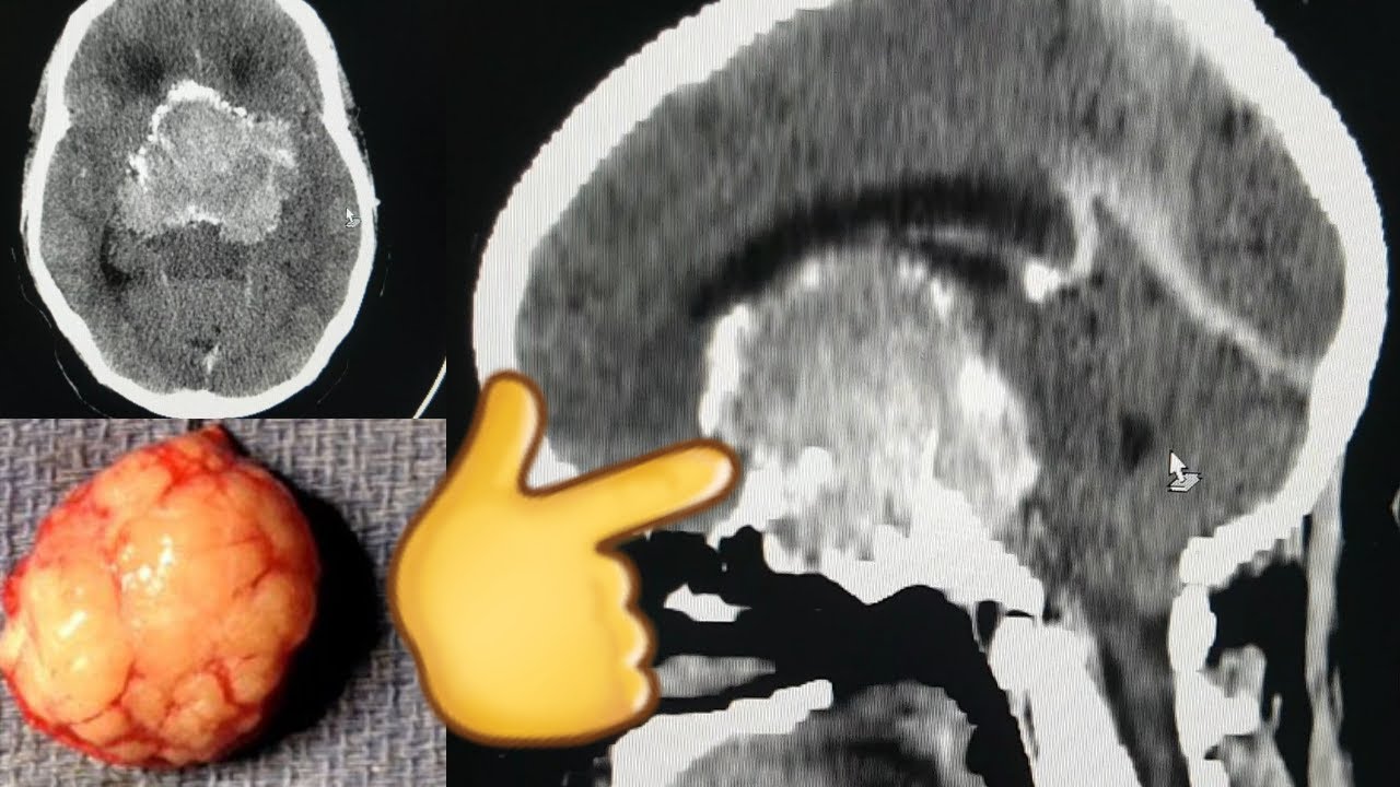
 brain ct scan for female 50 years old patent with history of chronic headache and vision loss for 6 months .
there is large lobulated heterogeneous hyper dense sellar and supra sellar with intense encasement on post contrast and having peripheral nodular calcification,encased the vessel of circle of Willis and destruction of the sellar walls,the sphenoid body ,greater and lesser sphenoid wings bilaterally and the clivus with lytic infiltrative lesions .............
Impression
large intra and suprasellar mass secondary to ?meningioma with adjacent bone destruction and extension to adjacent structures,mild obstructive hydrocephalus ,encasement of the vessels of the circle of Willis and signs of raised ICP.
NB .pituitary macroadenoma can be possible differential.


0 Comments